Common Mistakes in Peptide Reconstitution: A Laboratory Prevention Guide
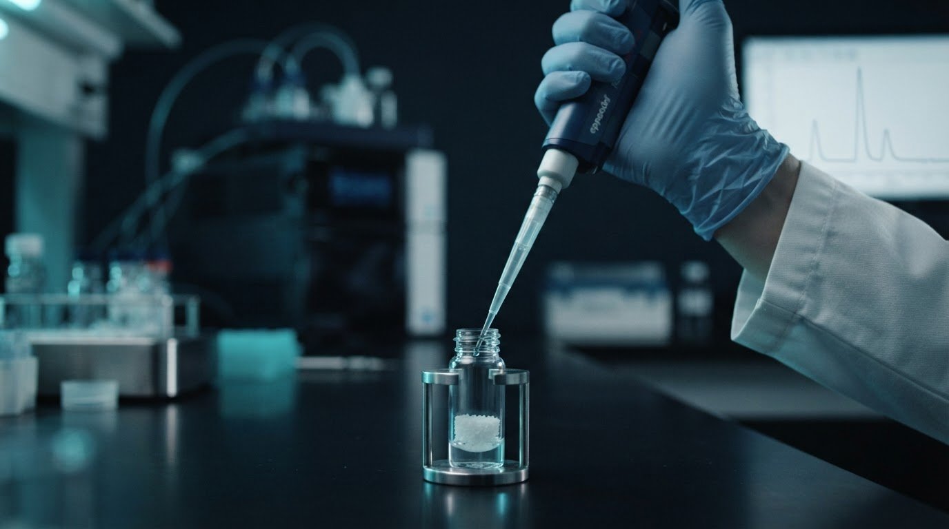
Precision in synthesis is useless without precision at the bench. Most peptide degradation in the laboratory does not stem from inherent chemical instability; it occurs the moment diluent hits the lyophilized cake. Few disruptions stall an assay faster than inspecting a freshly solubilized vial only to observe turbidity, flocculation, or persistent particulate. While researchers often suspect compound synthesis when analytical runs fail, the true drivers of structural compromise are almost always common mistakes in peptide reconstitution, spanning aggressive mechanical shear to severe solvent mismatches.
Even high-purity research materials degrade rapidly under uncontrolled hydrodynamic turbulence or incorrect pH exposure. You can prevent this failure point entirely. By mastering precise, sequence-specific preparation protocols, you will safeguard delicate molecular structures, eliminate concentration errors, and preserve continuous solution stability across ongoing research workflows. Below, we break down the verified laboratory standards for thermal equilibration, solvent selection, and fluid dynamics required to execute flawless reconstitution every time.
Key Takeaways
- Prevent immediate structural denaturation by mastering controlled solvent delivery along the vial wall rather than direct impingement onto the lyophilized cake.
- Identify sequence-dependent solvent requirements to eliminate common mistakes in peptide reconstitution that cause rapid aggregation and pH-driven deamidation.
- Eliminate concentration discrepancies and assay variance by properly distinguishing total lyophilized cake mass from net peptide content during volumetric calculations.
- Protect reconstituted molecular chains against crystalline shear stress by replacing destructive freeze-thaw cycles with single-use aliquoting protocols.
- Establish rigorous thermal equilibration and post-solubilization visual inspection benchmarks to confirm baseline solution integrity prior to experimental use.
Biochemical Foundations of Reconstitution and the Mechanics of Peptide Degradation
Reconstitution transforms a fragile, freeze-dried matrix into an active liquid research solution. Understanding the underlying chemistry of peptide synthesis illuminates why these compounds arrive lyophilized. Lyophilization purges water to stabilize peptide backbones against hydrolytic cleavage. Yet, this process leaves an amorphous, highly reactive cake. When researchers introduce a diluent without respecting molecular thermodynamics, secondary structural motifs such as alpha-helices and beta-sheets denature instantly. Committing common mistakes in peptide reconstitution leads to immediate strand cleavage, macro-aggregation, and irreproducible baseline assay readings.
The Fragile Architecture of Lyophilized Peptide Cakes
Freeze-drying creates an ultra-porous, hygroscopic lattice. This structure preserves tertiary conformations in the dry state, but it remains hyper-reactive to atmospheric variables. Moisture is an immediate catalyst for hydrolytic breakdown. Exposing cold peptide containers to ambient room air draws immediate condensation directly onto the cake, causing localized solvation before controlled reconstitution even begins.
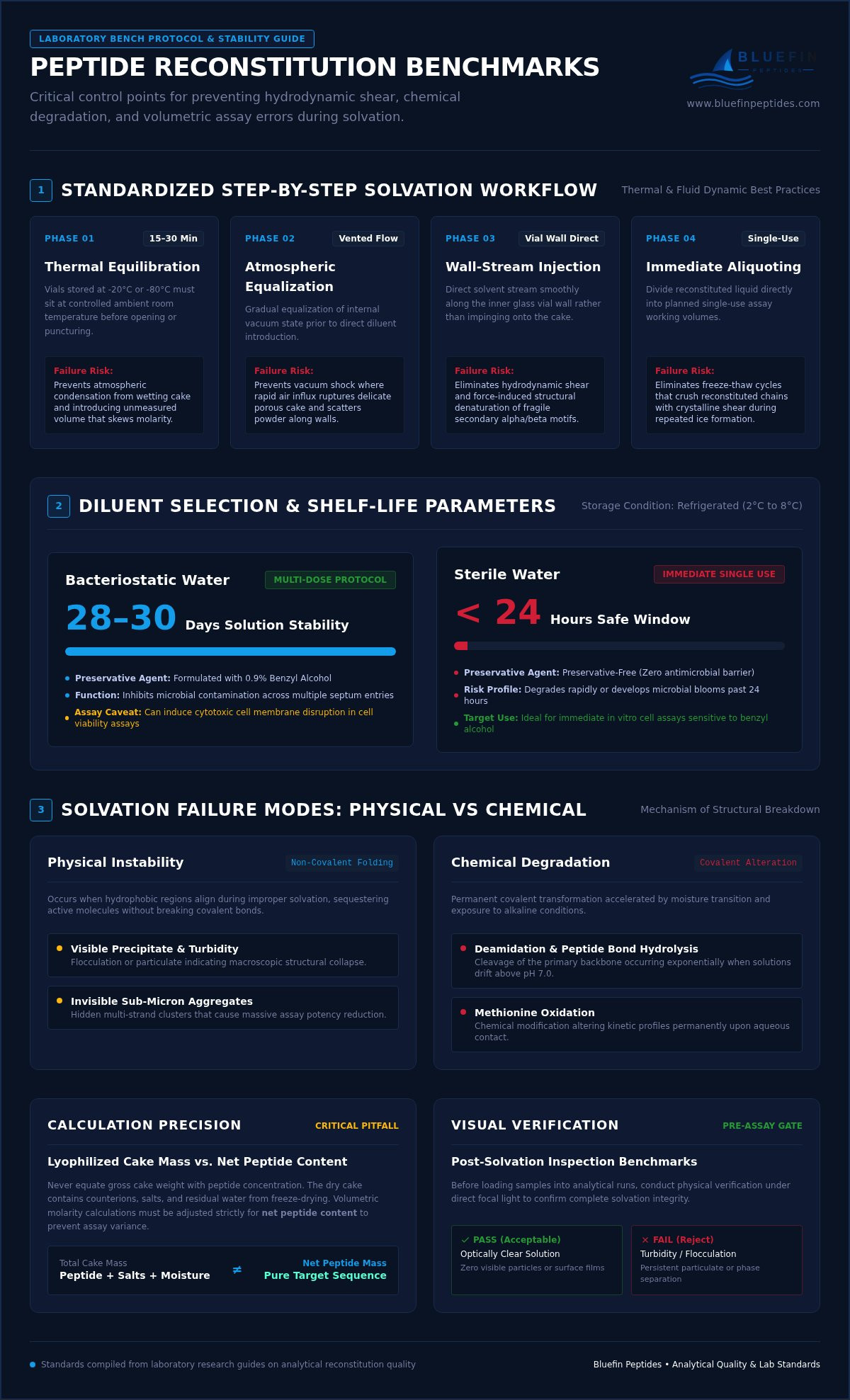
- Equilibrate thermal baselines: Vials stored at -20°C or -80°C must sit at controlled ambient room temperature for 15 to 30 minutes before puncturing or removing the septum.
- Prevent vacuum shock: Rapid air influx into an unvented vacuum vial ruptures the delicate lyophilized scaffold, scattering particles across the vial walls.
- Control relative humidity: Ambient condensation introduces unpredictable water volume, skewing molarity calculations before research protocols initiate.
Chemical vs Physical Degradation During Solvation
Solvation introduces two distinct failure modes: irreversible chemical alterations and physical aggregation. Physical instability arises when hydrophobic patches on folding peptide strands align, generating visible precipitate or invisible sub-micron aggregates. These aggregates sequester active molecules, causing dramatic drops in assay potency. In contrast, chemical degradation permanently alters the primary covalent structure through deamidation, peptide bond hydrolysis, or methionine oxidation. These reactions surge exponentially once compounds transition from dry states into aqueous media, especially when solutions drift above pH 7.0.
Flawless laboratory reconstitution requires starting with fully authenticated materials. Ensuring that compounds adhere to verified analytical quality standards for research peptides guarantees that issues observed post-solvation stem from handling variables rather than pre-existing batch synthesis impurities. Bypassing common mistakes in peptide reconstitution protects your sequence from irreversible degradation, preserving baseline kinetic profiles from the first aliquot to the last.
Diluent Selection Mistakes: Bacteriostatic Water, Sterile Saline, and pH Pitfalls
Selecting the wrong carrier fluid ranks among the most destructive common mistakes in peptide reconstitution. Every synthesized sequence possesses a distinct isoelectric point (pI) and hydropathy profile. Treating water as a universal solvent ignores fundamental thermodynamics. Without matching solvent polarity to peptide charge, researchers trigger rapid precipitation, phase separation, or catastrophic hydrolysis before the first trial run starts.
Bacteriostatic Water vs Sterile Water in Extended Assays
Bacteriostatic Water contains 0.9% benzyl alcohol, functioning as an antimicrobial preservative. This bacteriostatic additive maintains chemical stability across multi-dose research protocols, preserving reconstituted integrity for 28 to 30 days when stored at 2°C to 8°C. Preservative-free sterile water lacks this defense; solutions prepared with plain sterile water degrade or harbor microbial blooms within 24 hours under identical refrigeration.
However, benzyl alcohol is not universally benign. In cell-based viability assays, the preservative can induce cytotoxic cell membrane lysis, skewing experimental results. In spectrophotometric evaluations below 260 nm, benzyl alcohol creates dense optical interference that masks protein absorption peaks. Selecting between preserved and unpreserved matrices requires matching experimental timelines to downstream detection methods.
Solubility Screening for Hydrophobic and Basic Sequences
Net molecular charge dictates liquid solubility. Peptides containing basic residues, such as arginine, lysine, or histidine, frequently form opaque suspensions at neutral pH. Forcing basic compounds into solution requires a targeted drop in solvent pH:
- Basic peptide dissolution: Solubilize the lyophilized cake in a minimal volume of dilute acetic acid (0.1% to 1.0%) before bringing the solution up to final volume with sterile water.
- Acidic peptide dissolution: Compounds rich in aspartic or glutamic acid dissolve cleanly in slightly alkaline solutions; a dilute ammonium hydroxide solution (0.1%) creates the necessary ionic environment.
- Hydrophobic sequences: Peptides containing high ratios of leucine, isoleucine, or valine resist aqueous ingress entirely, requiring preliminary dissolution in pure dimethyl sulfoxide (DMSO) or acetonitrile before aqueous dilution.
Attempting to dissolve resistant sequences by flooding the vial with excess sterile water dilutes experimental molarity without resolving thermodynamic repulsion. This error leads to visible flocculation and uneven concentration gradients. Avoiding these common mistakes in peptide reconstitution requires aligning bench protocols with verified analytical data, such as the comprehensive batch documentation provided by Bluefin Peptides for experimental sequence validation.
Mechanical Errors During Reconstitution: Agitation, Shear Stress, and Pressure Traps
Mechanical trauma destroys molecular integrity just as definitively as chemical contamination. While solvent selection addresses thermodynamic stability, physical handling controls structural preservation. Peptide chains rely on fragile hydrogen bonding and hydrophobic interactions to maintain secondary folding. Applying uncontrolled kinetic energy fractures these weak stabilization forces. Overlooking physical fluid mechanics remains one of the most pervasive common mistakes in peptide reconstitution, transforming pristine research materials into biologically inactive debris.
The Science of Shear Stress and Why Shaking Destroys Peptides
Shaking or vortexing a reconstitution vial generates destructive hydrodynamic shear. As liquid accelerates violently across the glass interior, velocity gradients tear apart sensitive tertiary structures. This agitation forces atmospheric gas into the liquid matrix, creating persistent surface foam. Frothing is not benign effervescence. It signals extensive air-liquid interfacial denaturation, where unfolded hydrophobic peptide cores aggregate irreversibly along gas bubble surfaces.
Eliminating shear stress requires strict adherence to low-velocity handling principles:
- Abolish vortex mixing: Never subject reconstituted peptide solutions to mechanical vortex mixers or aggressive reciprocal shaking.
- Implement horizontal rolling: Submerge the lower vial base and roll it slowly between gloved palms to initiate gentle fluid circulation.
- Utilize passive dissolution: Allow the diluent to wet the matrix spontaneously; unhurried diffusion preserves conformation far better than forced motion.
Pressure Equalization and Wall-Directed Diluent Injection
Most laboratory peptide vials are sealed under a partial negative pressure vacuum to stabilize the dry matrix. Puncturing the septum without regulating plunger resistance allows this vacuum to yank the syringe barrel forward instantly. This produces a high-velocity fluid jet that blasts directly into the lyophilized cake, causing immediate mechanical fracturing. Highly sensitive, sequence-dense compounds like the retatrutide research peptide demand rigorous flow control to prevent localized structural shear.
Mitigate internal vial pressure by following a standardized fluid introduction sequence:
- Maintain manual plunger tension: Hold the syringe plunger firmly between your thumb and index finger before penetrating the rubber stopper, actively resisting the inward vacuum pull.
- Angle the needle bevel: Direct the needle tip toward the internal glass sidewall at a 45-degree angle, well above the lyophilized matrix.
- Execute slow laminar release: Depress the plunger gradually, allowing the diluent to trickle down the glass perimeter and flood the base softly from beneath.
- Equalize internal atmosphere: Withdraw residual negative pressure using a sterile, empty syringe barrel or venting needle before storing the solubilized compound.
Eliminating these common mistakes in peptide reconstitution safeguards molecular geometry, ensuring experimental assays deliver true baseline readouts rather than artifacts of physical trauma.
Calculation, Aliquoting, and Storage Pitfalls That Compromise Research Assays
Assay failures frequently occur long after physical solvation appears complete. Miscalculating final concentration, storing solutions in improper plasticware, or subjecting reconstituted aliquots to repetitive thermal cycles silently invalidates an experimental series. These post-solvation missteps represent some of the most insidious common mistakes in peptide reconstitution because the resulting solution appears visually pristine despite massive degradation or active peptide loss.
Math and Volume Pitfalls: Concentration vs Syringe Measurement
Lyophilized vial labels indicate total dry compound mass, not reconstituting liquid capacity. A persistent laboratory error involves conflating vial mass with final target concentration. Establishing precise working stock requires a deliberate formula:
Concentration (µg/µL) = [Total Mass (mg) × 1,000] ÷ Diluent Volume (µL)
Crucially, researchers must account for net peptide content. Lyophilized cakes contain counter-ions, such as trifluoroacetate or acetate salts, along with residual moisture. A 10 mg cake rarely yields 10 mg of pure peptide mass. Standard analytical data on a Certificate of Analysis (COA) documents net peptide percentages, which often range from 70% to 85%. Failing to adjust solvent volume against documented batch purity skews molarity calculations and distorts dose-response kinetics across downstream assays.
Aliquoting Strategies and Thermal Degradation Prevention
Liquid peptides face rapid surface adsorption and atmospheric degradation. Sequences adhere nonspecifically to standard polystyrene; laboratories must exclusively utilize low-retention, sterile polypropylene microcentrifuge tubes to prevent severe analyte depletion. Excessive vial headspace also accelerates oxidative pathways by exposing the liquid meniscus to ambient oxygen.
Thermal stability requires strict aliquoting discipline to prevent crystalline shearing:
- Eliminate freeze-thaw cycles: As water freezes, expanding ice crystals exert intense shear stress that cleaves peptide backbones. Never freeze a master stock vial more than once.
- Subdivide immediately: Partition freshly reconstituted solutions into single-use aliquots calibrated to daily assay volumes.
- Observe thermal parameters: Maintain short-term working aliquots at 2°C to 8°C for up to 30 days when formulated with bacteriostatic preservation. Store unpreserved solutions at -20°C or -80°C for extended research.
- Monitor multi-target solutions: Complex formulations like the GHK-Cu BPC-157 TB-500 blend contain distinct peptide chains with unique solubility limits; immediate subdivision prevents differential precipitation over time.
Avoiding these common mistakes in peptide reconstitution preserves the molecular stability verified during synthesis. To establish reliable experimental baselines with verified purity, source analytical-grade peptides from Bluefin Peptides for your laboratory workflows.
Standardized Laboratory Reconstitution Protocol and Quality Verification
Reproducibility across laboratory assays requires standardizing every manual variable. Inconsistent technique introduces experimental noise that obscures genuine analytical outcomes. Establishing an institutional Standard Operating Procedure (SOP) eliminates technician variance, ensuring that baseline kinetic readings reflect compound biology rather than handling discrepancies. Systematic quality verification guarantees that common mistakes in peptide reconstitution don't corrupt downstream analytical workflows.
The Complete Step-by-Step Laboratory Reconstitution SOP
Follow this validated operational sequence for consistent reconstitution across all in vitro research workflows:
- Sanitize the workspace: Decontaminate the laminar flow hood surface with 70% isopropanol. Allow the airflow to stabilize for five minutes before staging supplies.
- Equilibrate thermal baselines: Remove the research vial from storage (-20°C or -80°C) and allow it to sit sealed at ambient temperature (20°C to 25°C) for 20 to 30 minutes.
- Inspect the lyophilized matrix: Visually assess the dry cake through the borosilicate glass. The cake should appear uniform and intact, showing no signs of pre-solvation collapse or discoloration.
- Aseptically prepare the septum: Remove the flip-off cap and wipe the chlorobutyl rubber stopper with a sterile alcohol swab; allow the surface to air-dry completely.
- Introduce the verified diluent: Draw the exact calculated solvent volume into a sterile syringe. Puncture the septum at an angle, directing the needle bevel toward the inner glass wall while resisting vacuum pull. Depress the plunger slowly.
- Facilitate passive wetting: Allow the vial to rest undisturbed for two to five minutes. Slowly swirl or roll the vial horizontally between your palms until the compound dissolves entirely. Hydrophobically dense compounds like the tirzepatide research peptide require extended passive wetting to dissolve without localized aggregation.
- Conduct optical verification: Hold the vial against both light and dark backgrounds. The reconstituted solution must be transparent and free of suspended fibrils, haze, or particulate matter.
- Document and store: Record reconstitution date, diluent lot number, calculated molarity, and storage coordinates in the laboratory information management system (LIMS). Aliquot immediately into sterile polypropylene tubes.
Analytical Quality Control and Research Assurance
Flawless bench technique cannot compensate for subpar reagent synthesis. Preventing common mistakes in peptide reconstitution starts before diluent ever touches glass: it begins with independent batch verification. Baseline analytical confirmation requires ≥99% HPLC purity testing to confirm the absence of truncated sequences, deletion variants, or synthesis byproducts that compromise solubility.
Equally critical is mass spectrometry documentation. Electrospray ionization mass spectrometry (ESI-MS) confirms exact target molecular mass, proving sequence authenticity prior to solvent addition. When your laboratory sources validated compounds from the Bluefin Peptides high-purity laboratory catalog, US-stocked inventory backed by comprehensive batch Certificates of Analysis ensures that experimental anomalies trace back to assay mechanics, never synthesis integrity.
Mastering Bench Precision for Reliable Experimental Outcomes
Rigorous science leaves no room for procedural ambiguity. Flawless reconstitution bridges the gap between raw chemical synthesis and reproducible analytical data. By aligning solvent polarity with sequence hydropathy, eliminating destructive hydrodynamic shear, and replacing repetitive freeze-thaw cycles with single-use aliquoting, laboratories systematically avoid the common mistakes in peptide reconstitution that invalidate sensitive assays.
Even perfect technique demands an uncompromised starting point. Analytical integrity begins with verified purity. When your research requires uncompromising baseline reagents, procure HPLC-verified compounds for your laboratory assays at Bluefin Peptides. With ≥99% HPLC purity verification, full mass spectrometry batch confirmation, and rapid delivery from US-stocked storage facilities, you can approach every in vitro protocol with complete experimental confidence.
Frequently Asked Questions
Why did my peptide solution turn cloudy or milky after adding diluent?
Cloudiness indicates insoluble aggregation or incomplete solvation caused by an isoelectric point mismatch. If the carrier fluid matches the compound's net neutral charge, electrostatic repulsion vanishes, forcing molecules to clump. You can sometimes resolve slight haziness with gentle rolling. If turbidity persists, the sequence requires a modified pH environment, such as dilute acetic acid or ammonium hydroxide, rather than neutral water.
Can I reconstitute research peptides with standard sterile saline instead of water?
Using standard 0.9% sodium chloride is generally not recommended for initial lyophilized cake dissolution. The high ionic strength of saline can initiate salting-out effects, causing immediate precipitation in sensitive compounds. It's best practice to dissolve the cake in sterile or bacteriostatic water first. Once the peptide is fully in solution, you can dilute it into isotonic saline buffers for specific analytical workflows.
What happens to peptide structure if a research vial is vigorously shaken?
Vigorous agitation generates intense hydrodynamic shear stress that ruptures fragile secondary structures. The resulting foam forces peptide chains to unfold along air-liquid interfaces, exposing hydrophobic cores that aggregate into biologically inert clusters. Shaking stands out among the most destructive common mistakes in peptide reconstitution. Always use slow, horizontal palm rolling to protect delicate tertiary folding.
How many times can an aliquoted peptide solution be safely frozen and thawed?
Reconstituted peptide solutions should undergo zero repeated freeze-thaw cycles. Freezing creates sharp, microscopic ice crystals that shear peptide backbones, while thawing drives localized aggregation. Submitting a solution to more than three cycles causes massive structural degradation. Laboratories must partition freshly solubilized material into single-use polypropylene aliquots immediately after reconstitution.
Does bacteriostatic water alter the pH balance of reconstituted research compounds?
Bacteriostatic water typically exhibits a slightly acidic pH ranging between 4.5 and 7.0 due to dissolved atmospheric carbon dioxide and 0.9% benzyl alcohol. Most synthetic peptides remain stable within a mildly acidic range of 4.0 to 6.0, making this carrier ideal. However, basic peptides requiring neutral or alkaline environments may precipitate if the slight acidity isn't balanced with a dedicated buffer.
How do I equalize vial vacuum pressure safely without contaminating the lyophilized powder?
Equalize internal vacuum pressure by regulating syringe resistance manually during solvent delivery. Puncture the septum while gripping the plunger firmly to prevent the negative pressure from sucking the diluent in violently. Alternatively, insert an open, sterile needle through the stopper to vent the headspace, ensuring air flows solely through an attached 0.22-micron sterile syringe filter to prevent airborne particulate contamination.
How long do reconstituted peptides maintain chemical stability under refrigeration at 4°C?
Peptides reconstituted with bacteriostatic water maintain analytical stability for approximately 28 to 30 days when held at 2°C to 8°C. Solutions prepared with preservative-free sterile water degrade within 24 hours due to hydrolytic pathways and bacterial growth risk. Avoiding common mistakes in peptide reconstitution ensures your solution reaches its full projected bench lifespan without premature molecular breakdown.
For laboratory research use only. Not for human or veterinary use. This content is educational and does not constitute medical, dosing, or usage guidance.
Browse the BLUEFIN catalog