Establishing Peptide Stability in Solution Through Solubility
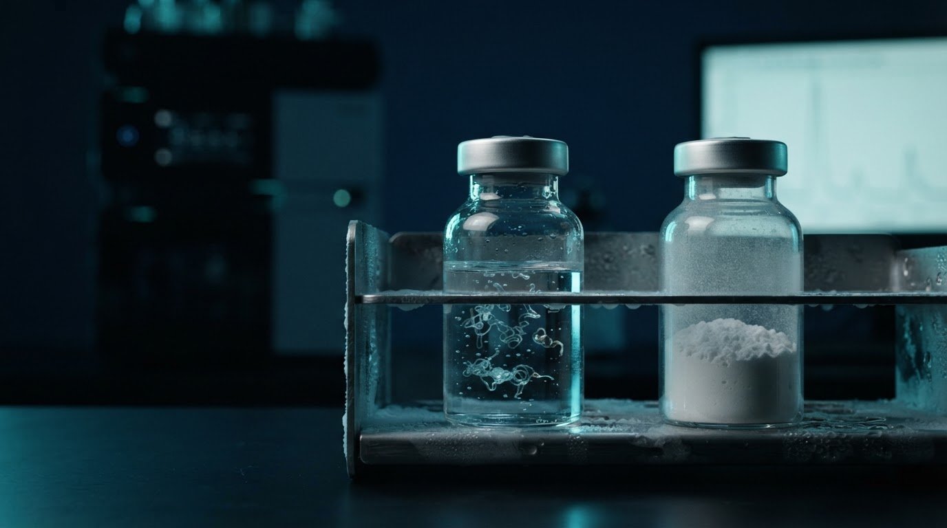
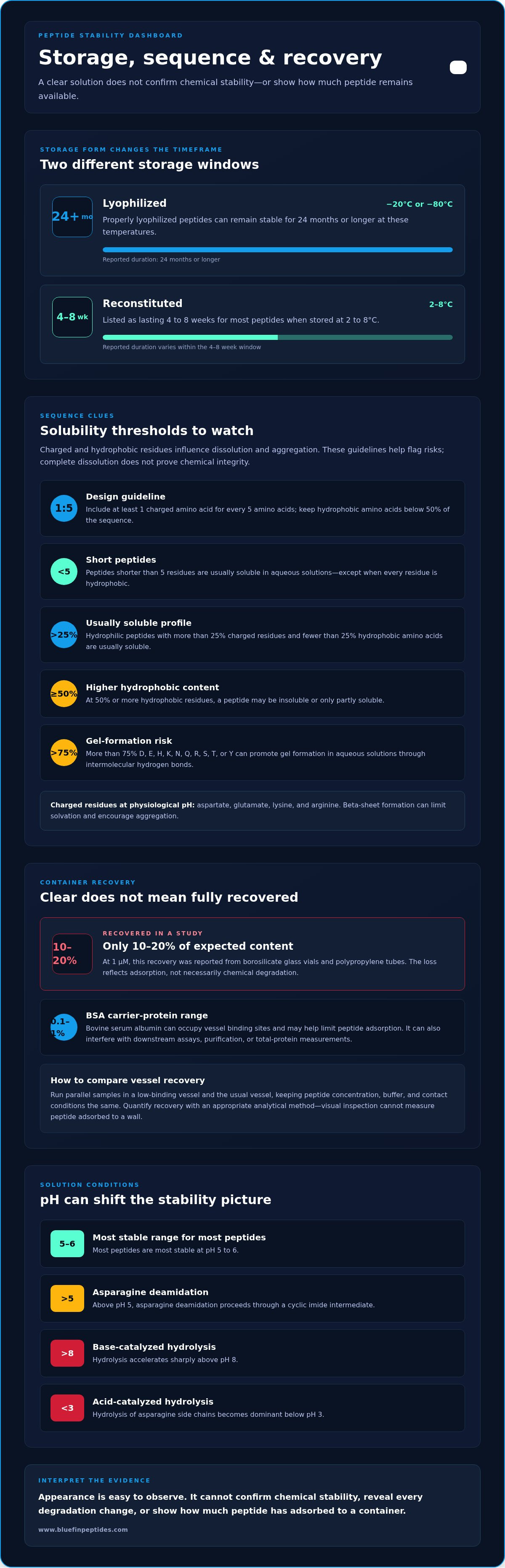
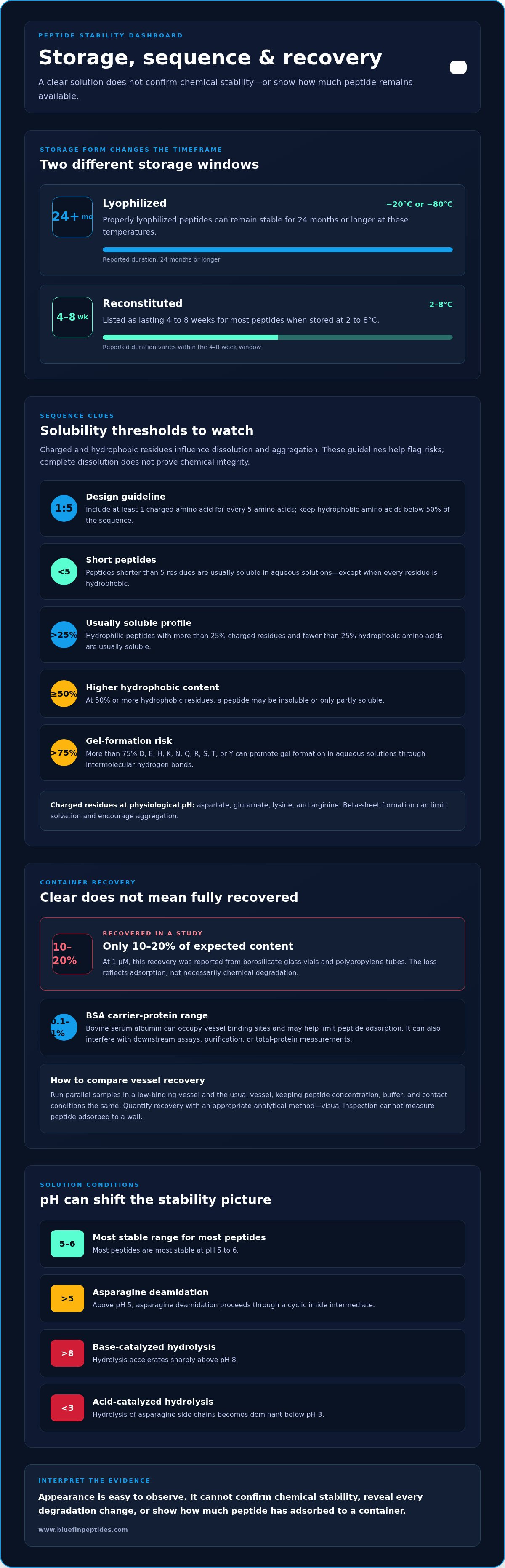
A peptide solution can look clear and unchanged while its chemical integrity has declined; visual inspection cannot confirm stability. Establishing peptide stability in solution depends on factors such as sequence, solvent, pH, container interactions, temperature, and time after reconstitution. Properly lyophilized peptides stored at −20°C or −80°C can remain stable for 24 months or longer, while reconstituted peptides stored at 2 to 8°C are listed as lasting 4 to 8 weeks for most peptides.
Appearance is easy to observe, but it cannot reveal every chemical change or confirm that a peptide retains its intended properties. This distinction matters when interpreting a solution and its storage history.
This article explains how amino acid composition affects aqueous solubility, why peptides can adsorb to glass or plastic, and how solvents, containers, and reconstitution influence stability. It also compares lyophilized and reconstituted forms, including the effects of temperature and freeze-thaw handling, so you can assess evidence without relying on visual checks alone.
Key Takeaways
- Use amino acid composition to anticipate aqueous solubility: charged and hydrophobic residues influence whether a peptide dissolves fully or forms aggregates.
- Choose containers and handling conditions with surface adsorption in mind; carrier proteins such as BSA may help reduce peptide loss.
- Match the solvent to the peptide’s properties, since a solvent that supports dissolution can still affect downstream experiments or solution behavior.
- Distinguish lyophilized storage from reconstituted storage, and limit temperature changes and repeated freeze-thaw cycles to help preserve peptide integrity.
- A clear solution is not proof of chemical stability; visual inspection cannot identify every form of degradation.
Amino acid composition and aqueous solubility
Peptide stability in aqueous solution depends on sequence, pH, temperature, and the tendency to dissolve or aggregate. Hydrophobic and charged amino acids influence solubility, while oxidation-sensitive residues and hydrolysis-prone bonds affect chemical integrity. These factors interact: complete dissolution does not establish that a peptide has avoided chemical change.
At physiological pH, aspartate, glutamate, lysine, and arginine have charged side chains. A design guideline is to include at least one charged amino acid for every five amino acids and keep hydrophobic amino acids below 50% of the sequence. Peptides shorter than 5 residues are usually soluble in aqueous solutions, except when every residue is hydrophobic. Hydrophilic peptides with more than 25% charged residues and fewer than 25% hydrophobic amino acids are usually soluble. At 50% or more hydrophobic residues, a peptide may be insoluble or only partly soluble. For establishing peptide stability in solution, these sequence features help identify solubility risks before selecting conditions.
Sequence patterns can also promote gel formation or aggregation. Peptides containing more than 75% of D, E, H, K, N, Q, R, S, T, or Y can form gels in aqueous solutions through intermolecular hydrogen bonds. Beta-sheet formation can limit solvation and encourage aggregation. Changing repeated residue patterns with conservative substitutions may help limit beta-sheet formation. A single substitution, such as replacing alanine with glycine, or adding polar amino acids to a terminus may also improve solubility.
Cysteine, methionine, and tryptophan are oxidation-sensitive residues. Multiple instances can complicate high-purity peptide production because oxidation or side reactions may occur. Design alternatives include norleucine as a substitute for methionine and serine as a less reactive replacement for cysteine. Unnatural amino acids offer another design tool. Introducing β-amino acids into neurotensin-derived peptide 53 extended its half-life to 32 hours. Replacing the N-terminal arginine with β-arginine in neurotensin-derived peptide 54 resulted in a half-life greater than 7 days. These examples concern plasma half-life, not aqueous solubility.
To minimize oxidation, deamidation, hydrolysis, and aggregation, evaluate residue composition alongside solution conditions. Amber vials or foil-wrapped containers are recommended for solutions containing tryptophan, methionine, or tyrosine. Most peptides are most stable at pH 5 to 6, although asparagine deamidation above pH 5 proceeds through a cyclic imide intermediate. Base-catalyzed hydrolysis accelerates sharply above pH 8, while acid-catalyzed hydrolysis of asparagine side chains becomes dominant below pH 3. A sound approach to establishing peptide stability in solution considers composition, pH, and storage conditions together.
Low-binding containers and carrier proteins
Peptide recovery depends on more than whether a solution appears clear. Cationic peptides can bind to container surfaces, reducing the amount available for an experiment. In a study at 1 μM, only 10 to 20% of the expected peptide content was recovered from borosilicate glass vials and polypropylene tubes. This loss reflects adsorption, not necessarily chemical degradation. A sample can therefore contain less freely dissolved peptide than its prepared concentration suggests.
Low-binding containers can reduce surface interactions, but their benefit depends on the peptide and experimental setup. Compare recovery in the intended container using the same peptide concentration, buffer, and contact conditions. Run parallel samples in a low-binding vessel and the usual vessel, then quantify peptide recovery with an appropriate analytical method. Visual inspection cannot measure peptide adsorbed to a wall. This comparison helps distinguish surface loss from changes caused by the solution itself.
Carrier proteins provide another way to limit adsorption. Bovine serum albumin, or BSA, can occupy binding sites on vessel surfaces before a peptide does. The recommended concentration range is 0.1 to 1%. This approach can be useful for dilute peptide preparations, where surface-bound material can represent a meaningful portion of the sample. However, BSA adds protein to the solution. That can interfere with downstream assays, purification, or measurements that detect total protein. Select a carrier only when its presence is compatible with the experimental readout.
Container choice and carrier use address physical recovery, while sequence design can target other stability problems. An N-terminal asparagine can present a synthesis challenge because its protecting group may be difficult to remove at that position. Possible design responses include removing or replacing the residue, or adding one residue to lengthen the peptide. These changes alter the sequence, so they are design decisions, not universal handling fixes. They also do not prevent adsorption to glass or plastic.
Protein fusion is a separate strategy for improving the behavior of some peptides. Attaching a peptide to a larger carrier protein can shield exposed hydrophobic regions and change how the fused construct interacts with the solution and surfaces. The fusion partner also changes molecular size, structure, and assay behavior. If an experiment requires the isolated peptide, the fusion may need a cleavage site and a separation step. That additional processing can cause recovery losses, so compare the fused construct with the intended final peptide rather than assuming fusion alone ensures stability.
For a research workflow, match the approach to the problem: test vessel recovery for adsorption, consider BSA when compatible, and reserve sequence changes or protein fusion for design-level needs. research peptide options can help identify a peptide for a defined laboratory study.
Organic solvents for peptide dissolution
Organic solvents can help dissolve sequences that resist water, especially when hydrophobic residues dominate. Dimethyl sulfoxide (DMSO), dimethylformamide (DMF), and acetonitrile are used in peptide work. They are not interchangeable: the choice depends on the peptide, the next experimental step, and whether the solvent affects the assay. Dissolution is only the first check. A peptide that forms a clear stock in DMSO may precipitate after dilution into an aqueous buffer.
Start with the sequence and intended final solution. A strongly hydrophobic peptide may need an organic solvent to disperse, while a charged sequence may dissolve directly in aqueous buffer. Consult the peptide’s documented handling conditions, then test a small sample before preparing the full working solution. This limits unnecessary solvent exposure that could alter conformation, interfere with analysis, or prove incompatible with the experiment.
Dilution is a critical transition. Add the organic-solvent stock gradually to the aqueous phase while mixing, rather than adding the full volume at once. Local differences in solvent composition can trigger aggregation or precipitation before the solution is uniform. For example, a peptide dissolved in DMSO may form visible particles if added rapidly to a water-based buffer. Examine the mixture after each addition, and use an analytical method to compare recovery if appearance alone is inconclusive.
Solvent compatibility also shapes interpretation. DMSO can affect assays involving enzymes, membranes, or protein interactions, so include a solvent-only control with the same final solvent content. DMF or acetonitrile may also interfere with downstream measurements, depending on the method. Keep the solvent proportion consistent across control and peptide samples. If the solvent-only control changes the assay response, that signal cannot be confidently attributed to the peptide.
Distinguish solubility from chemical stability. A solvent can keep a peptide dispersed without preventing oxidation, deamidation, hydrolysis, or other sequence-dependent changes. Conversely, precipitation can reduce recoverable peptide without proving chemical degradation. Record the solvent identity, dilution order, buffer composition, and appearance at each stage. For sequences containing oxidation-sensitive residues such as methionine or tryptophan, limit unnecessary light exposure with an amber vial or foil wrapping. These records help distinguish solvent-driven behavior from later changes in the solution.
Adsorption to glass and plastic
Peptide disappearance from a sample does not automatically indicate chemical degradation. A peptide may bind to the container wall, leaving less freely available for the assay target. In a study of cationic peptides at 1 μM, recovery from borosilicate glass vials and polypropylene tubes was only 10 to 20% of the expected content. The measured result can therefore reflect both peptide integrity and losses to the vessel surface.
This effect matters especially when interpreting dilute samples. A fixed amount of adsorbed peptide represents a larger share of a small sample than of a concentrated one. Surface chemistry matters too: glass and polypropylene differ, so one vessel type cannot represent every container. Compare the actual materials used in the workflow, including the storage vial and the tube used during analysis.
Separate surface loss from degradation with a controlled recovery comparison. Prepare matching peptide samples and hold the peptide, buffer, concentration, and contact time constant, changing only the vessel material. Measure recovered peptide with a suitable analytical method rather than relying on clarity or visible residue. Include a reference sample with minimal contact with vessel surfaces. A lower result in one container suggests a recovery issue, but does not by itself identify the binding mechanism.
For example, if a sample produces a lower assay signal after storage in polypropylene than in borosilicate glass, repeat the comparison with fresh aliquots and consistent handling. Record the sample concentration, container material, buffer, and time before measurement. If the difference persists, assess whether the assay measures total peptide or only freely available peptide. This distinction affects interpretation: surface-bound material may be unavailable to the assay without having undergone chemical change.
Low-binding vessels can be evaluated as a practical way to limit surface-associated losses. Carrier proteins are another option in compatible experiments; BSA at 0.1 to 1% is recommended to reduce adsorption. However, added protein can interfere with assays that measure protein content or require purified peptide. Select the approach around the experimental target and its readout. A vessel comparison, analytical recovery measurement, and clear handling record provide stronger evidence than appearance alone. They also help distinguish adsorption-related loss from degradation during solution storage.
Lyophilized versus reconstituted peptide stability
Lyophilized peptides stored at −20°C or −80°C can remain stable for 24 months or longer. Reconstitution changes the storage picture: most peptides are listed as lasting 4 to 8 weeks at 2 to 8°C, or 24 to 72 hours at room temperature.
Formulation Stability Requirements
FDA and ICH guidance inform how stability evidence is generated for peptide drug formulations. Research peptides are not the same as approved drug products. Formulation stability assessments use defined conditions and evaluate whether critical quality attributes remain within established specifications during storage. This distinction matters because results for lyophilized material cannot automatically establish the stability of its reconstituted solution.
Study records should identify the formulation, container, storage conditions, and analytical endpoints. A peptide’s sequence and formulation affect which changes matter, including loss of intact peptide, aggregation, or altered appearance. Evidence must therefore match the material and storage state being evaluated.
Effects of pH and Buffer Choice
pH changes reaction rates and can shift the degradation pathways affecting peptide residues. Most peptides are described as most stable at pH 5 to 6. Asparagine deamidation above pH 5 proceeds through a cyclic imide intermediate, while base-catalyzed hydrolysis accelerates sharply above pH 8. Below pH 3, acid-catalyzed hydrolysis of asparagine side chains becomes dominant.
Buffer choice helps maintain the selected pH, but the buffer must also suit the peptide and analytical method. Compare the same formulation across controlled pH conditions, and track changes using a stability-indicating measurement. Temperature matters too: reaction rates in solution chemistry roughly double for every 10°C increase.
Analytical Methods for Stability Measurement
Analytical methods measure changes that visual inspection cannot establish. HPLC can track intact peptide and related purity changes, while mass spectrometry can help identify molecular-mass changes associated with chemical modification. Combining separation with mass analysis helps distinguish a declining parent-peptide signal from the appearance of potential degradation products.
Record the initial result as a baseline, then use the same validated method for later samples. Include a suitable reference and consistent sample preparation. Turbidity or precipitation can indicate physical change, but a clear sample does not prove that the peptide remains chemically intact.
Designing Solution Stability Studies
Design the study around the intended storage state: lyophilized, reconstituted, or aliquoted. Take a baseline measurement immediately after preparation, then select later time points to capture early changes and longer-term trends. Keep peptide concentration, container, buffer, light exposure, and temperature consistent across comparison groups.
Reconstituted aliquots stored at −80°C are listed as lasting months to a year; at −20°C, weeks to months. Each freeze-thaw cycle can reduce bioactivity by 5 to 15%, depending on the peptide, so use separate aliquots to limit repeated thawing. Bacteriostatic water contains 0.9% benzyl alcohol and inhibits bacterial growth across repeated aseptic withdrawals over a 28-day in-use period, but it does not prevent chemical degradation.
Bluefin Peptides: Retatrutide Research Peptide
Bluefin Peptides offers Retatrutide Research Peptide for laboratory research requiring a defined peptide source. Each peptide is ≥99% HPLC-verified, with mass-spectrometry confirmation and a batch-specific certificate of analysis (COA). These records support review of identity and purity as researchers plan their own solution-stability studies; they do not replace study-specific measurements of a reconstituted sample.
Choose a research peptide according to the study’s target and experimental design. Bluefin Peptides also offers Tirzepatide Research Peptide, BPC-157 / TB-500 Research Blend, and Ipamorelin / CJC-1295 Research Blend. Review the available product information when selecting an offering for laboratory use.
Visit Bluefin Peptides online to explore its research peptide offerings.
Set Up Your Next Stability Study
Turn stability observations into a defined research plan. Choose the peptide, intended solution conditions, and analytical endpoint before comparing samples. This alignment makes changes easier to interpret and helps identify which variables deserve closer study. Establishing peptide stability in solution is an experimental question, answered with evidence from the specific peptide and conditions under evaluation.
For laboratory research, Bluefin Peptides offers Retatrutide Research Peptide, Tirzepatide Research Peptide, and BPC-157 / TB-500 Research Blend. These offerings provide options to consider when selecting research material for a planned study. Keep the material’s documentation and your study records together, so observations remain traceable to the sample and conditions tested.
Explore Bluefin Peptides’ research offerings as you plan your next laboratory study. A clear experimental question is a strong starting point for reliable interpretation.
Frequently Asked Questions
Can I tell visually if a peptide solution has degraded?
No. A clear solution does not establish that the peptide remains chemically intact, because changes can occur without visible color or particles. Cloudiness can signal aggregation or incomplete dissolution, but does not identify the cause. Treat appearance as an observation, not a stability result. Compare stored and fresh samples using an analytical method that separates intact peptide from related components.
Does bacteriostatic water prevent chemical degradation?
No. Bacteriostatic water contains 0.9% benzyl alcohol, which inhibits bacterial growth during repeated aseptic withdrawals over a 28-day in-use period. It does not prevent chemical reactions that alter a peptide’s structure. For example, oxidation, deamidation, and hydrolysis are distinct from microbial growth. Choosing bacteriostatic water therefore does not replace appropriate storage or chemical stability measurements.
How long does a reconstituted peptide last in the refrigerator?
Reconstituted peptides stored at 2 to 8°C are listed as lasting 4 to 8 weeks for most peptides. The appropriate interpretation depends on the specific peptide, its solution conditions, and the study’s stability criteria. Record the reconstitution date and storage conditions for each sample. A refrigerator timeline is a reference for planning, not evidence that a particular sample retained its original composition.
How many freeze-thaw cycles can a peptide tolerate?
Each freeze-thaw cycle can reduce bioactivity by 5 to 15%, depending on the peptide, so repeated cycling can affect experimental results. There is no single cycle count that applies equally to every sequence. Track each thaw and use separate aliquots to limit repeated temperature changes. Thaw aliquots on ice and use them within hours of thawing, as handling time also affects the sample.
How to Enhance the Pharmacokinetic Stability of Peptides?
Pharmacokinetic stability can be improved through structural modifications that reduce enzymatic breakdown or alter clearance. Examples include changing backbone chemistry or attaching a fatty-acid chain, with each modification also potentially affecting activity and distribution. In one reported comparison, a thioamide-containing peptide had a 50-fold increase in plasma half-life versus its amide-containing counterpart. Plasma half-life describes behavior in the body, not stability in a laboratory solution.
Why do peptides stick to glass and plastic tubes?
Peptides can interact with vessel surfaces, reducing the amount available in solution. The effect depends on peptide properties, concentration, and material, so glass and plastic may produce different recoveries. For example, dilute cationic peptide samples can show substantial surface-associated loss. Comparing recovery of the same sample in different vessels can help identify adsorption as a cause of low measured peptide, rather than assuming degradation.
What temperature and storage conditions preserve peptides in solution?
For reconstituted peptides, 2 to 8°C is associated with a listed duration of 4 to 8 weeks for most peptides; room-temperature storage is listed as 24 to 72 hours. Protect solutions containing tryptophan, methionine, or tyrosine from light with amber vials or foil wrapping. Temperature, light exposure, and solution composition should be recorded together when interpreting stability measurements.
How does peptide sequence and structure influence solution stability?
Sequence influences both dissolution and chemical susceptibility. Hydrophobic residues can limit aqueous solubility, while cysteine, methionine, and tryptophan can be vulnerable to oxidation. Asparagine can undergo deamidation, and repeated hydrogen-bonding patterns can encourage aggregation. As a result, two peptides in the same buffer may behave differently. Assess the sequence alongside solution conditions, and measure chemical changes rather than inferring stability from solubility alone.
For laboratory research use only. Not for human or veterinary use. This content is educational and does not constitute medical, dosing, or usage guidance.
Browse the BLUEFIN catalog